2-Methoxyestradiol (2-MeOE2) Derivatives Inhibit Orai1 Store-Operated Calcium Entry in Neuroinflammation
Copy code to add this image to your page
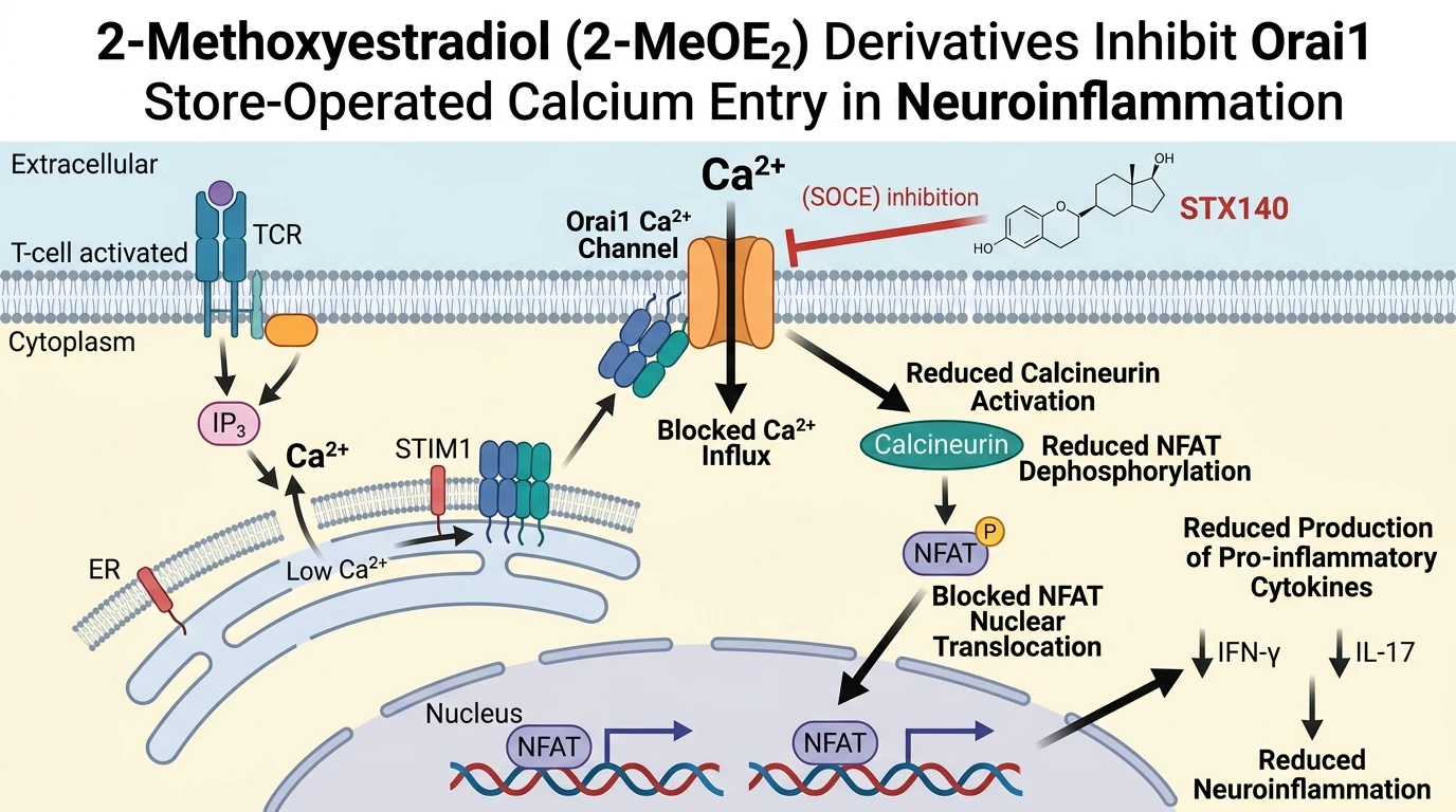
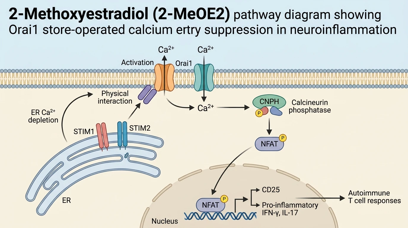
Store-operated calcium entry (SOCE) serves as a principal Ca2+ influx pathway in non-excitable cells, particularly T lymphocytes. Upon T cell receptor (TCR) engagement, phospholipase C-gamma catalyzes the hydrolysis of phosphatidylinositol 4,5-bisphosphate into inositol 1,4,5-trisphosphate (IP3) and diacylglycerol. Binding of IP3 to its receptors on the membrane of the endoplasmic reticulum (ER) triggers luminal Ca2+ depletion. ER-resident transmembrane sensors, STIM1 and STIM2, detect this loss of Ca2+ ions and translocate to ER-plasma membrane junctions. There, STIM molecules interact physically with Orai1 pore-forming proteins, opening the Ca2+ selective channel. The resulting influx of extracellular Ca2+ sustains cytosolic calcium elevations required for long-term downstream signal transduction in activated immune cells.
Intracellular Ca2+ elevations directly activate the calmodulin-dependent phosphatase calcineurin. Dephosphorylation of nuclear factor of activated T cells (NFAT) by calcineurin unmasks nuclear localization signals, allowing NFAT to translocate into the cell nucleus. Within the nuclear compartment, NFAT acts alongside co-activators to induce transcription of inflammatory genes, including interleukin-2 receptor alpha chain (CD25), interferon-gamma (IFN-γ), and interleukin-17 (IL-17). Disruption of this signaling cascade attenuates autoreactive T cell expansion, differentiation, and inflammatory cytokine output. Consequently, targeting Orai1-mediated SOCE represents a compelling strategy for mitigating autoimmune neuroinflammation.
Chemical modification of the natural endogenous estrogen metabolite 2-Methoxyestradiol (2-MeOE2) yielded the bis-sulfamoylated derivative 2-Methoxyestradiol-3,17-O,O-bis-sulfamate (STX140). While inhibition of store-operated calcium entry was historically examined in non-immune models, STX140 demonstrates marked potency and selectivity for SOCE suppression in T cells. In vitro spectrofluorometric assays utilizing fura-2 loaded T cells establish that STX140 blocks store-operated Ca2+ influx with half-maximal inhibitory concentrations (IC50) ranging from 20 to 27 µM. Channel blockade occurs rapidly upon compound exposure without depleting internal Ca2+ stores prior to activation.
Functional downstream consequences of SOCE blockade by STX140 are evident across multiple cellular models. In rat myelin basic protein-reactive T cells (TMBP) and human peripheral blood mononuclear cells (PBMCs), STX140 inhibits TCR-dependent activation in a concentration-dependent manner. Surface expression of CD25 drops significantly following treatment. Furthermore, secretion of signature pro-inflammatory cytokines, specifically IFN-γ and IL-17, is dramatically suppressed in both rodent and human lymphocyte preparations. These findings confirm that pharmacological SOCE inhibition by STX140 directly blunts the activation phenotype of encephalitogenic T lymphocytes.
The mechanistic independence of STX140 from classical steroid sulfatase (STS) inhibition was confirmed through comparative studies. Parent compound 2-Methoxyestradiol (2-MeOE2) and the highly specific pure STS inhibitor Irosustat were evaluated alongside STX140 in identical experimental systems. Neither parent 2ME2 nor Irosustat reproduced the robust inhibition of SOCE observed with STX140. Furthermore, STS inhibition alone failed to suppress T cell cytokine output or CD25 upregulation. These comparative data demonstrate that the anti-inflammatory activity of STX140 relies upon direct Orai1 channel modulation rather than STS enzyme inhibition or classical estrogen receptor activation.
In vivo efficacy was validated using experimental autoimmune encephalomyelitis (EAE) models in Lewis rats. Daily intraperitoneal administration of STX140 at 10 mg/kg/day achieved complete protection against clinical paralysis in both active immunization and adoptive transfer EAE paradigms. Histopathological evaluation of central nervous system (CNS) tissues revealed a near-total absence of immune cell infiltration into the spinal cord parenchyma. Neuroinflammatory biomarkers were markedly attenuated. In contrast, control animals treated with vehicle or parent 2-Methoxyestradiol (2-MeOE2) developed severe ascending paralysis and extensive neuroinflammation. These in vivo results underscore the therapeutic utility of SOCE blockade in autoimmune central nervous system disorders.
Therapeutic Evaluation and SOCE Blockade Characterization
Copy code to add this image to your page
2-Methoxyestradiol (2-MeOE2) derivatives such as STX140 act as potent inhibitors of store-operated Ca2+ entry (SOCE) mediated by Orai1 channels. Blocking intracellular Ca2+ influx prevents calcineurin-dependent nuclear translocation of NFAT in T lymphocytes. Consequently, downstream expression of activation markers (CD25) and release of encephalitogenic cytokines (IFN-γ and IL-17) are suppressed. This targeted channel inhibition operates independently of steroid sulfatase (STS) activity, providing robust neuroprotective and anti-inflammatory efficacy in experimental models.
STX140 Suppresses Lymphocyte SOCE and Cytokine Release
In vitro evaluation confirms that STX140 targets Ca2+ homeostasis in T lymphocytes. Calcium spectrofluorometry demonstrated that STX140 inhibits store-operated Ca2+ entry with an IC50 of 20–27 µM. Channel blockade occurred independently of intracellular ER calcium store depletion. Downstream signaling ceased rapidly. In rat TMBP cells and human PBMCs, TCR-stimulated upregulation of CD25 was markedly reduced. Production of IFN-γ and IL-17 decreased in a dose-dependent fashion. Neither parent 2-Methoxyestradiol (2-MeOE2) nor pure STS inhibitor Irosustat exerted comparable inhibitory effects under identical assay conditions. The anti-inflammatory potency of STX140 thus stems directly from Orai1 channel interference rather than steroid sulfatase inhibition.
Parenteral STX140 Attenuates Neuroinflammation in Rat EAE
Daily intraperitoneal administration of STX140 at 10 mg/kg/day completely prevented clinical symptoms in Lewis rat EAE models. Both active myelin-induced and adoptive transfer EAE cohorts exhibited zero motor paralysis. Histology confirmed absence of leukocyte infiltration within CNS spinal cord sections. Microglial activation was reduced. In contrast, parent 2-Methoxyestradiol (2-MeOE2) failed to alter disease progression or tissue pathology at equivalent doses. These empirical observations establish that STX140 provides complete functional protection against encephalitogenic T cell-driven neuroinflammation.