GSK-3 Inhibitor IX inhibitor (BIO) Modulates Canonical Wnt Signaling to Maintain Stem Cell Pluripotency
Copy code to add this image to your page
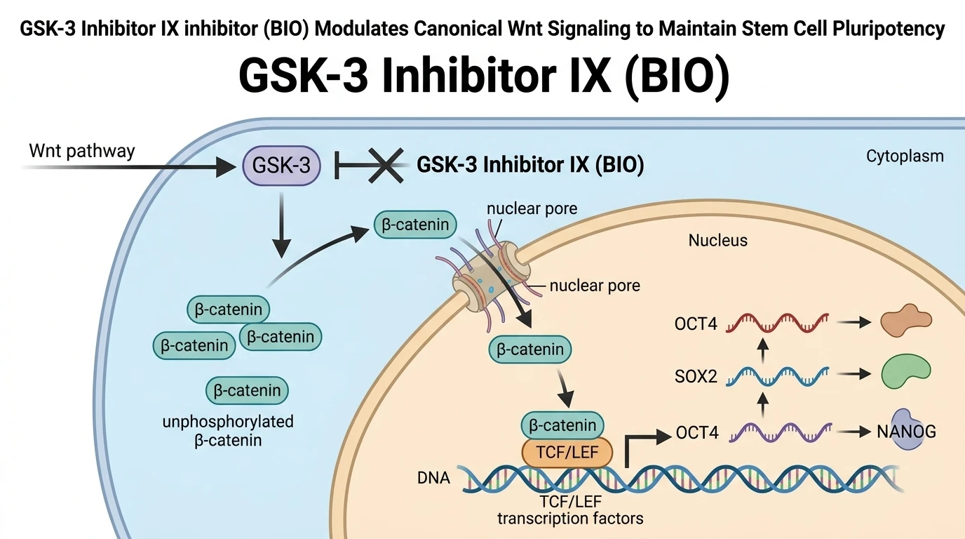
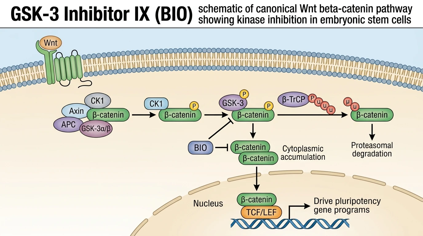
Glycogen synthase kinase-3 (GSK-3) functions as a central serine/threonine kinase regulating diverse metabolic, developmental, and transcriptional pathways. The enzyme exists as two cellular isoforms, GSK-3α and GSK-3β, which share high sequence homology within their catalytic domains. Under basal conditions, GSK-3 maintains active constitutive phosphorylation of intracellular target substrates. Within the canonical Wnt/β-catenin signaling cascade, GSK-3 forms an integral component of a cytosolic destruction complex alongside axin, adenomatous polyposis coli (APC), and casein kinase 1 (CK1). Active GSK-3 phosphorylates specific amino-terminal serine and threonine residues (Ser33, Ser37, Thr41) on β-catenin. This phosphorylation event flags β-catenin for recognition by the E3 ubiquitin ligase subunit β-TrCP. Rapid ubiquitination and subsequent 26S proteasomal degradation maintain cytoplasmic β-catenin at minimal baseline levels.
Pharmacological interception of this constitutive turnover is achieved using GSK-3 Inhibitor IX (BIO, 6-bromoindirubin-3'-oxime). BIO acts as a cell-permeable, highly potent, and ATP-competitive inhibitor of both GSK-3α and GSK-3β isoforms. By direct binding to the ATP-binding pocket of the kinase domain, the GSK-3 Inhibitor IX inhibitor prevents terminal phosphate transfer to primed protein substrates. Inhibition occurs at low nanomolar biochemical concentrations (IC50 ≈ 5 nM), demonstrating high selectivity relative to closely related cyclin-dependent kinases such as CDK1 and CDK5. When tested in cell culture models, ATP-competitive engagement destabilizes the operational assembly of the destruction complex without requiring extracellular Wnt receptor ligation.
Blockade of GSK-3 catalytic activity halts the amino-terminal phosphorylation cascade of β-catenin. Unphosphorylated β-catenin evades recognition by the β-TrCP ubiquitin ligase machinery. Consequently, the protein accumulates rapidly within the cytosolic compartment. Upon reaching critical threshold concentrations, stabilized β-catenin translocates across the nuclear membrane. Within the nucleus, β-catenin displaces transcriptional corepressors, such as Groucho/TLE, from T-cell factor/lymphoid enhancer factor (TCF/LEF) DNA-binding proteins. The resulting β-catenin-TCF/LEF transcriptionally active complex recruits coactivators CBP/p300 and BRG1. This complex directly initiates transcription of target genes governing cellular self-renewal and pluripotency networks, including OCT4, SOX2, and NANOG.
In human and mouse embryonic stem cell (ESC) cultures, small-molecule GSK-3 inhibition serves as an essential tool for maintaining undifferentiated cell fates. Traditional pluripotency maintenance required complex co-culture systems with feeder layers or exogenous cytokine support, such as leukemia inhibitory factor (LIF) or basic fibroblast growth factor (bFGF). Application of BIO preserves self-renewal capacity in defined, serum-free conditions by enforcing sustained endogenous Wnt target gene activation. Under tested in vitro assay conditions, prolonged treatment with BIO maintains pluripotency marker expression and colonies display characteristic undifferentiated morphology. However, the precise phenotypic outcomes depend heavily on dosage tuning, as excessive GSK-3 inhibition can trigger differentiation toward specific lineage lineages depending on the cellular context.
Pharmacological Mechanisms and Cellular Fate Regulation
Copy code to add this image to your page
GSK-3 Inhibitor IX (BIO) acts as a potent, cell-permeable, ATP-competitive inhibitor of GSK-3α and GSK-3β. Kinase inhibition blocks phosphorylation of β-catenin within the destruction complex. Unphosphorylated β-catenin escapes proteasomal degradation, accumulates in the cytosol, and translocates into the nucleus. Nuclear β-catenin binds TCF/LEF transcription factors, driving the transcription of self-renewal and pluripotency genes in embryonic stem cells.
ATP-Competitive Engagement of GSK-3 Isoforms in Vitro
Kinase domain profiling confirms that the GSK-3 Inhibitor IX inhibitor occupies the canonical ATP-binding cleft of GSK-3α and GSK-3β with high affinity. Hydrogen bonding with the hinge region stabilizes the inhibitor within the hydrophobic binding pocket. Phosphorylation of downstream substrates ceases within minutes of application. The selectivity margin over related cyclin-dependent kinases remains high under standardized in vitro assay conditions. This targeted kinase blockade prevents prime-dependent substrate processing without disrupting upstream signaling nodes.
Stabilization of Cytoplasmic β-Catenin and Translocation Dynamics
Inhibition of GSK-3 catalytic activity blocks N-terminal phosphorylation of β-catenin. Unphosphorylated β-catenin avoids recognition by β-TrCP ubiquitin ligases. Cytoplasmic degradation rates drop sharply. Consequently, unphosphorylated β-catenin accumulates in the cytosol before nuclear entry. In human and mouse embryonic stem cell models, this accumulation directly correlates with enhanced transcription at TCF/LEF consensus elements.
Maintenance of Pluripotency Markers in Embryonic Stem Models
Targeted application of GSK-3 Inhibitor IX (BIO) sustains pluripotency gene networks in embryonic stem cells. Marker expression levels of OCT4, SOX2, and NANOG remain elevated over extended passages in feeder-free conditions. Cellular viability remains intact across tested dosage ranges. These observations confirm that chemical stabilization of canonical Wnt signaling bypasses ligand dependence, supporting defined stem cell self-renewal.