Ammonium ferric citrate Suppresses FATP1-Mediated Fatty Acid Efflux Across Blood-Brain Barrier
Copy code to add this image to your page
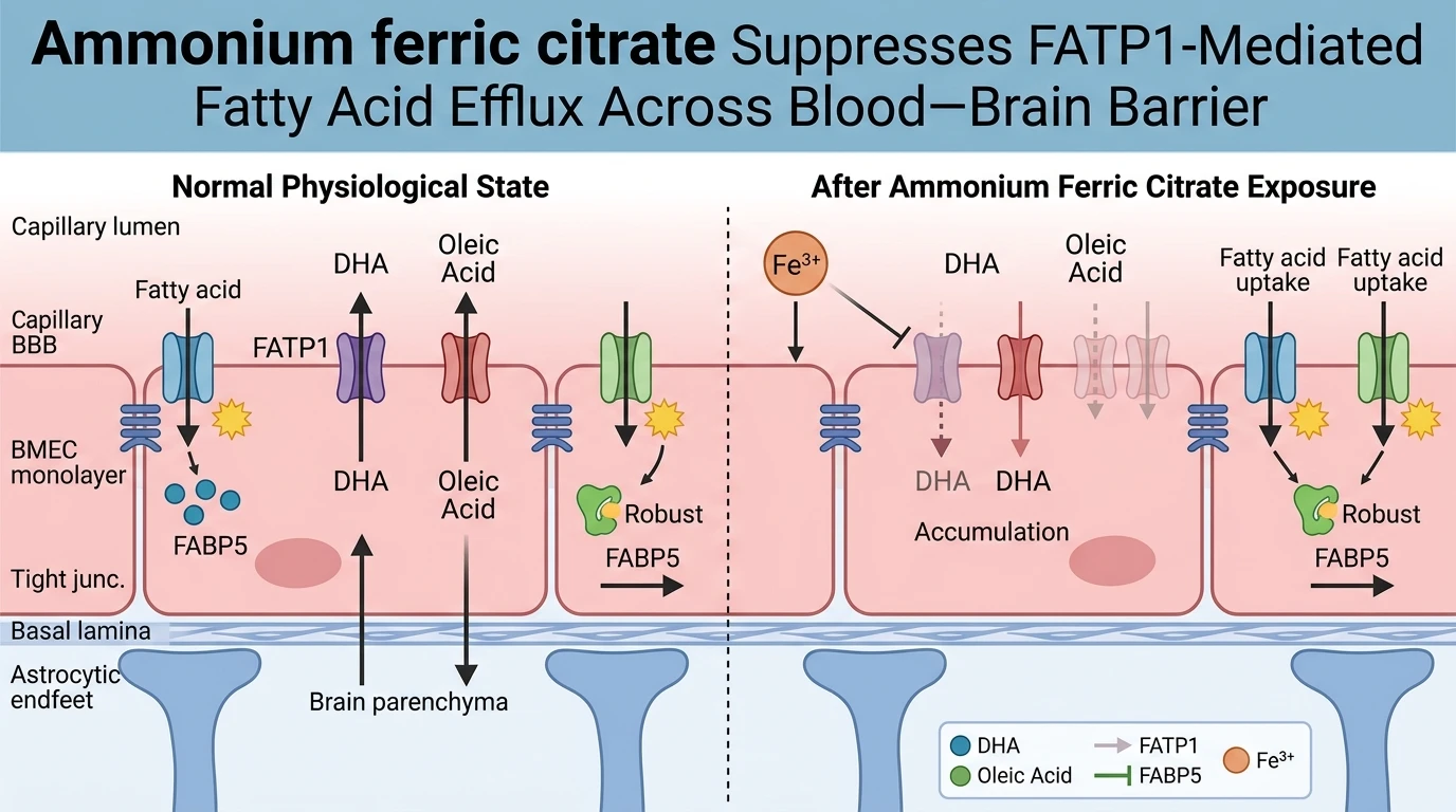
Brain microvascular endothelial cells forming the blood-brain barrier regulate the entry and exit of essential lipids required for central nervous system homeostasis. Among these lipids, docosahexaenoic acid represents a critical polyunsaturated fatty acid necessary for neuronal membrane fluidity, synaptic plasticity, and anti-inflammatory signaling. Because neural tissue lacks significant de novo synthesis capability for long-chain polyunsaturated fatty acids, brain tissue depends almost entirely on transport across the blood-brain barrier from systemic circulation. Fatty acid transport protein 1, encoded by the SLC27A1 gene, functions as a primary membrane-bound translocase facilitating fatty acid movement across endothelial boundaries. Intracellular iron homeostasis plays a major regulatory role in managing metabolic enzymes and transporter turnover within these vascular cells. In vitro investigations utilizing human immortalised brain microvascular endothelial cells (hCMEC/D3) demonstrate that exposure to Ammonium ferric citrate induces cellular iron overload, which alters FATP1 expression and function through distinct post-translational mechanics.
Quantitative analysis of endothelial cellular responses following exposure to Ammonium ferric citrate reveals a selective downregulatory effect on FATP1 protein abundance. Treatment with ferric ammonium citrate decreases FATP1 protein levels in a concentration-dependent manner without modifying SLC27A1 mRNA transcription. Reverse transcription quantitative PCR assays confirm stable transcript levels despite marked depletion of the mature FATP1 protein detected via immunoblotting. This dissociation between gene transcription and steady-state protein levels points toward a post-translational degradation mechanism or altered protein translation efficiency triggered by elevated intracellular labile iron pools. The preservation of transcript stability indicates that iron toxicity does not produce generalized transcriptional repression, but rather engages targeted proteolytic degradation or protein destabilization pathways selective for FATP1.
Functional transport assays using radiolabeled substrates elucidate the direct physiological consequences of FATP1 protein loss. In hCMEC/D3 monolayers treated with Ammonium ferric citrate, cellular efflux of both 3H-oleic acid and 14C-docosahexaenoic acid decreases significantly compared to vehicle-treated controls. FATP1 has traditionally been characterized primarily as a fatty acid uptake translocase, but these empirical transport metrics highlight its essential contribution to directed cellular efflux across endothelial membranes. Notably, the reduction in fatty acid transport is strictly compartmentalized to outward transport mechanisms. Inward transport kinetics for 3H-oleic acid and 14C-docosahexaenoic acid remain fully intact across tested experimental conditions. This preservation of fatty acid entry indicates that apical membrane uptake components operate independently of the iron-sensitive FATP1 degradation cascade.
Specificity assays further demonstrate that iron-induced downregulation is restricted to particular transport nodes rather than causing global endothelial dysfunction. Fatty acid binding protein 5, an intracellular lipid chaperone co-expressed in brain microvascular endothelial cells, shows no alteration in expression levels or functional binding capacity following ferric ammonium citrate administration. Western blot analyses and ligand-binding assays confirm that FABP5 protein abundance and lipid-handling capacity remain steady under identical iron-loading protocols. Furthermore, cell viability assays verify that the observed transport deficits occur at non-cytotoxic concentrations, ruling out non-specific membrane damage or necrotic cell death as drivers of altered efflux kinetics.
These molecular findings provide a potential physiological framework for understanding lipid metabolic disturbances observed in neurodegenerative disorders. Elevated brain iron accumulation is a recognized pathological feature of Alzheimer's disease, occurring alongside pronounced reductions in cortical docosahexaenoic acid levels. The demonstrated suppression of FATP1-mediated DHA efflux by intracellular iron overload offers a mechanistic connection between dysregulated iron homeostasis and compromised brain lipid profiles. By selectively suppressing FATP1 protein abundance, excessive iron accumulation at the vascular interface impairs the trans-endothelial trafficking of essential polyunsaturated fatty acids required for neuronal maintenance. Continued investigation into the precise proteolytic pathways governing iron-dependent FATP1 degradation may clarify therapeutic targets aimed at preserving vascular lipid transport in neurodegenerative states.
Endothelial FATP1 Suppression and BBB Fatty Acid Dynamics
Copy code to add this image to your page
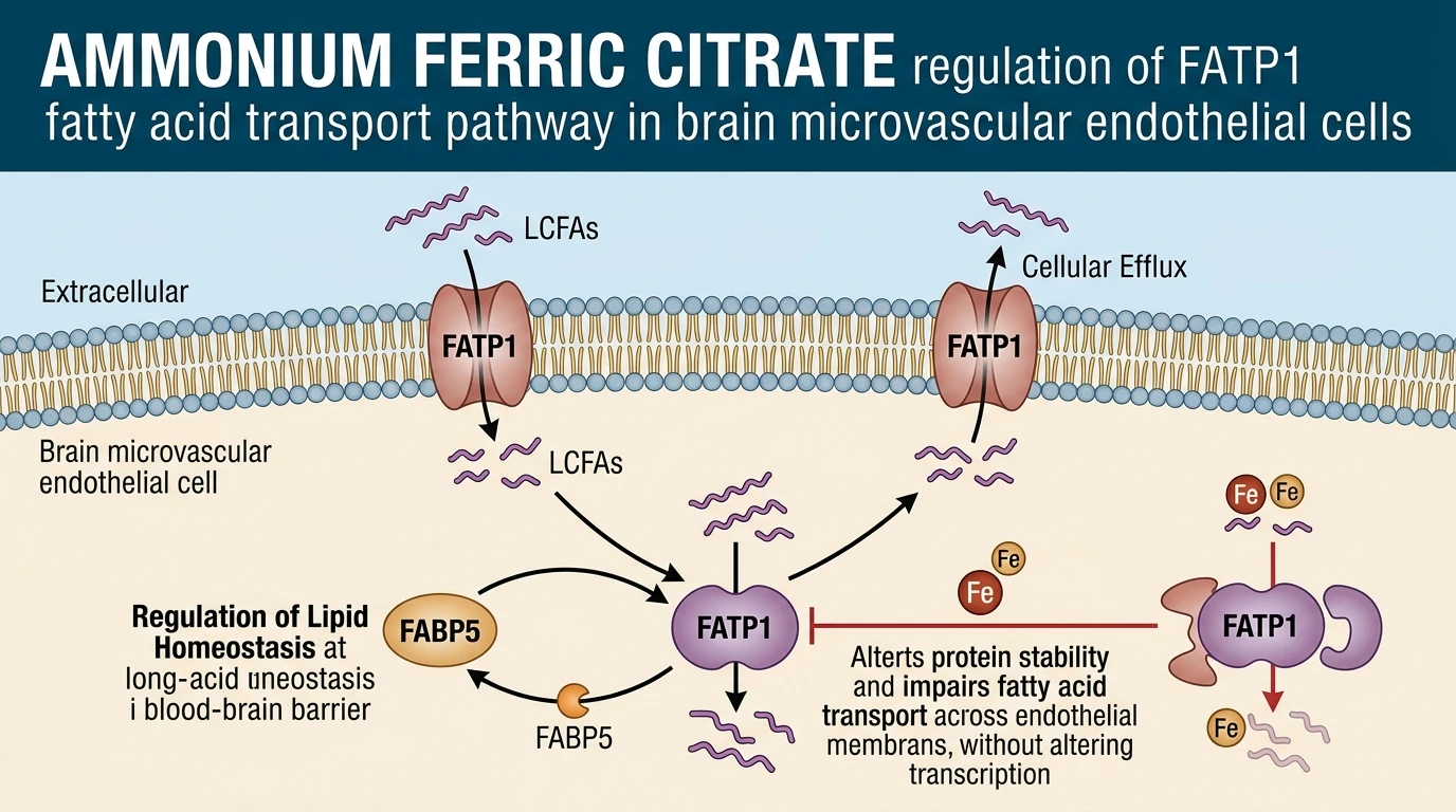
Ammonium ferric citrate induces endothelial iron overload, which selectively downregulates FATP1 protein abundance without modifying its transcript levels. This post-translational reduction decreases FATP1-mediated cellular efflux of polyunsaturated fatty acids, including docosahexaenoic acid (DHA) and oleic acid, across brain microvascular endothelial cells. Cellular fatty acid uptake and FABP5 expression remain unaffected under these conditions. Consequently, excess iron impairs FATP1-dependent DHA transport across the blood-brain barrier, providing a mechanistic explanation for diminished brain DHA concentrations in neurodegenerative pathologies.
Iron Overload Selective Downregulation of Endothelial FATP1 Abundance
Exposure to Ammonium ferric citrate selectively reduces FATP1 protein expression in human brain microvascular endothelial cells. Steady-state transcript levels remain entirely unaltered. Real-time quantitative PCR revealed identical SLC27A1 mRNA copy numbers between iron-loaded and control hCMEC/D3 cultures. In contrast, Western blot quantification confirmed a 50% drop in FATP1 protein signal within 24 hours of exposure—a rapid post-translational clearance event. Labile iron pools appear to accelerate intracellular protein degradation pathways. This degradation pattern occurs without causing overt cellular toxicity. Endothelial monolayer integrity remained fully intact under tested in vitro assay conditions. Thus, iron overload directly targets FATP1 protein stability rather than transcription.
Attenuated Efflux Kinetics of Docosahexaenoic Acid Across Endothelium
Radiolabeled tracer studies demonstrate selective inhibition of lipid transport across endothelial boundaries. Outward efflux dropped significantly. Cellular export of 14C-docosahexaenoic acid decreased markedly following Ammonium ferric citrate treatment, whereas total lipid uptake kinetics remained completely unchanged. Parallel experiments measuring 3H-oleic acid confirmed an identical efflux-specific transport deficit. Cellular uptake mechanics remained intact. Intracellular chaperone levels were similarly unaffected; FABP5 protein abundance and ligand-binding parameters displayed no alteration under high iron conditions. Selective FATP1 depletion directly impairs fatty acid efflux. These observations highlight a distinct post-translational node sensitive to heavy metal dynamics.