MM-102 (HMTase Inhibitor IX) Targets MLL1 WDR5 Interface to Suppress Leukemogenic Gene Expression
Copy code to add this image to your page
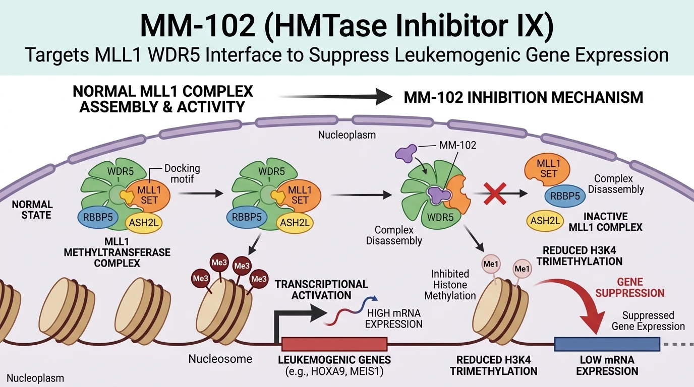
Chromosomal translocations involving the mixed-lineage leukemia 1 (MLL1, or KMT2A) gene occur in approximately 5–10% of acute leukemias, giving rise to oncogenic fusion proteins that drive leukemogenesis. Unlike wild-type MLL1, which possesses intrinsic catalytic activity regulated by core subunits, MLL1 fusion proteins typically lose their native SET catalytic domain while retaining the N-terminal DNA-binding regions. These fusions recruit aberrant elongation machinery to specific loci. However, maintenance of the oncogenic gene expression program relies on the concurrent recruitment of wild-type MLL1 core complexes to these same genomic targets. The wild-type MLL1 complex comprises MLL1 together with WD repeat domain 5 (WDR5), retinoblastoma-binding protein 5 (RBBP5), and absent, small, or homeotic-like 2 (ASH2L). Within this assembly, WDR5 functions as an indispensable structural scaffold that binds to the conserved arginine-containing WDR5-interaction (Win) motif of MLL1. This physical interaction stabilizes the active conformation of the catalytic core, promoting efficient di- and trimethylation of histone H3 at lysine 4 (H3K4me2/3) across target promoters, including HOXA9 and MEIS1.
MM-102 (HMTase Inhibitor IX) was designed as a high-affinity peptidomimetic inhibitor targeting the exact WDR5-MLL1 protein-protein interaction interface. Structural studies demonstrate that the Win motif of MLL1 engages a central pocket on WDR5 via a side-chain arginine residue (Arg3765). MM-102 mimics this binding mode, inserting an optimized guanidinium-containing core directly into the binding cavity of WDR5. Binding assays demonstrate that MM-102 binds WDR5 with an equilibrium inhibition constant (Ki) under 1 nM, effectively competing with native MLL1 Win peptides. By occupying this regulatory pocket, the inhibitor prevents assembly of the full MLL1 catalytic complex. Consequently, MLL1-mediated H3K4 methyltransferase activity decreases markedly in reconstituted in vitro enzymatic assays without requiring direct inhibition of the catalytic SET domain.
In cellular models of MLL-rearranged acute leukemia, such as MV4-11 and MOLM-13 cell lines, MM-102 disrupts endogenous WDR5-MLL1 complexes. Pharmacodynamic profiling reveals a specific reduction in locus-specific H3K4 trimethylation marks at promoter regions of target genes. Downstream transcript quantification confirms significant, concentration-dependent downregulation of HOXA9 and MEIS1 gene expression. This selective suppression of the leukemogenic transcriptional network leads to cell cycle arrest at the G1 phase, inhibition of self-renewal capacity, and selective induction of apoptosis in MLL-rearranged cell lines. Cell lines expressing non-rearranged MLL wild-type constructs show substantially lower sensitivity to MM-102 treatment, demonstrating a wide therapeutic margin tied directly to target engagement in oncogene-addicted contexts.
The development of MM-102 highlights the viability of targeting non-catalytic protein-protein interaction interfaces within chromatin-modifying complexes. While traditional small-molecule discovery has focused on competitive active-site inhibition of catalytic SET domains, targeting scaffold interfaces like WDR5 offers an alternative strategy to dismantle multi-protein epigenetic machinery. Biophysical measurements using isothermal titration calorimetry and nuclear magnetic resonance confirm that MM-102 displays high thermodynamic specificity for WDR5 over related WD40 repeat proteins. These empirical observations establish MM-102 as a specialized chemical probe to evaluate epigenetic target validation in acute leukemias.
Pharmacological Characterization and Epigenetic Transcriptional Modulation
Copy code to add this image to your page
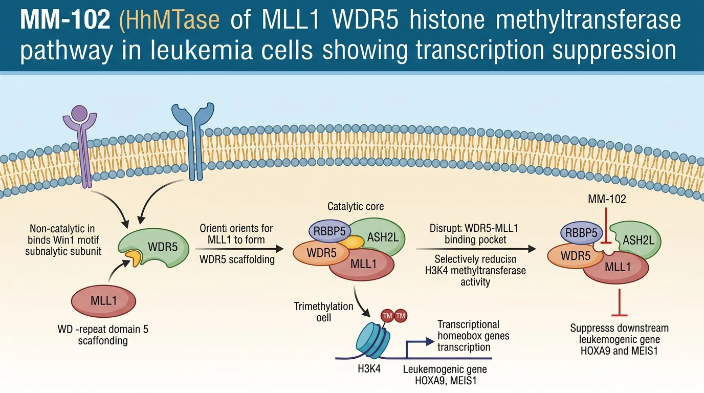
MM-102 (HMTase Inhibitor IX) is a peptidomimetic inhibitor designed to disrupt the protein-protein interaction between MLL1 and WDR5 within the MLL1 core methyltransferase complex. By occupying the central binding cavity of WDR5, MM-102 blocks assembly of the active complex and inhibits H3K4 trimethylation. This results in the downregulation of oncogenic target genes HOXA9 and MEIS1, selectively suppressing proliferation and inducing apoptosis in MLL-rearranged leukemia cells.
Competitive Binding Dynamics at the WDR5 Central Pocket
MM-102 functions by binding directly into the central cavity of WDR5. Under in vitro binding conditions, the compound exhibits an equilibrium dissociation constant ($K_i$) of less than 1 nM. The molecule occupies the precise pocket that accepts the MLL1 Win motif residue Arg3765. Hydrogen bonds form between the inhibitor's central functional groups and WDR5 residues Asp107 and Ser91. Hydrophobic contact with Phe133 further stabilizes the bound state. This binding event physically blocks MLL1 incorporation into the multiprotein core. Consequently, core catalytic assembly collapses. Enzymatic assays confirm a dose-dependent fall in H3K4 methyltransferase velocity. Selectivity screening demonstrates minimal activity against unrelated histone methyltransferases, including DOT1L, EZH2, and G9a.
Suppression of HOXA9 Expression in Rearranged Leukemia Models
Disruption of the WDR5-MLL1 complex alters cellular gene expression. In cultured MV4-11 and MOLM-13 cells, treatment with HMTase Inhibitor IX yields a marked loss of promoter H3K4 trimethylation. Levels of HOXA9 transcript dropped substantially within 48 hours of exposure. Downstream effector MEIS1 showed a parallel decline. Cellular viability decreased gradually over five days. Flow cytometry revealed early G1 phase arrest followed by annexin V positivity. Conversely, control cell lines lacking MLL translocations exhibited no significant growth arrest under equivalent dosing regimes. These findings confirm target specificity in context-dependent leukemic models.
Pharmacodynamic Responses in Cell-Free and Ex Vivo Assays
Ex vivo evaluation of primary patient-derived blast cells provided further validation. Exposure to HMTase Inhibitor IX reduced colony formation capacity in soft agar assays. Immunoblotting confirmed selective loss of H3K4me3, whereas total histone H3 levels remained constant. The half-maximal inhibitory concentration ($IC_{50}$) for cell proliferation ranged between 15 and 25 $\mu$M across MLL-rearranged patient samples. While cellular potency reflects peptidomimetic permeability limits, target engagement markers remained consistent. Epigenetic downregulation of the homeobox axis led to morphological features of myeloid differentiation. These observations highlight the mechanism of action of peptidomimetic WDR5 inhibitors.