Tucatinib (Irbinitinib, ONT-380, Arry-380) targeted HER2 kinase inhibition in preclinical models
Copy code to add this image to your page
Human epidermal growth factor receptor 2 (HER2/ERBB2) gene amplification drives aggressive tumor phenotypes in approximately twenty percent of human breast carcinomas. The small-molecule inhibitor Tucatinib (Irbinitinib, ONT-380, Arry-380) functions as a potent, ATP-competitive, highly selective inhibitor of the HER2 catalytic kinase domain. Unlike non-selective dual inhibitors such as lapatinib or neratinib, this agent displays low activity against epidermal growth factor receptor (EGFR/ERBB1) under tested in vitro kinase profiling conditions. High EGFR inhibition frequently induces dose-limiting cutaneous and gastrointestinal toxicities in patient populations. Consequently, target selectivity remains critical for establishing therapeutic windows during prolonged clinical administration schedules.
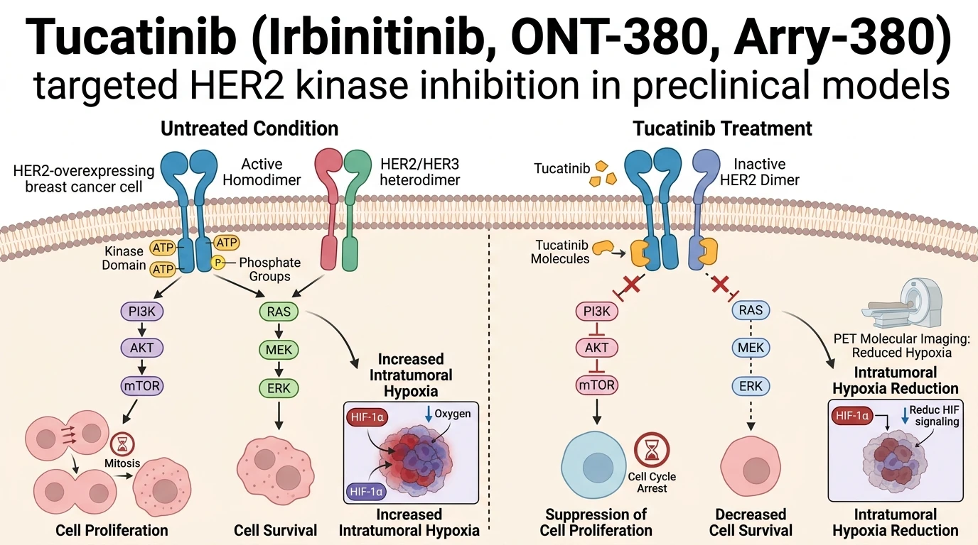
In preclinical studies utilizing the HER2-amplified BT474 breast cancer cell line and the BCM3472 patient-derived xenograft (PDX) model, oral administration of Tucatinib (Irbinitinib, ONT-380, Arry-380) yielded marked reductions in overall tumor burden. Investigators monitored these longitudinal pharmacodynamic shifts non-invasively through multi-tracer positron emission tomography (PET) imaging modalities. Cellular proliferation was evaluated using 2'-deoxy-2'-[18F]fluoro-L-thymidine ([18F]-FLT) PET scans alongside immunohistochemical Ki-67 scoring. Daily drug administration suppressed DNA synthesis pathways effectively. The tracer uptake fell within forty-eight hours post-treatment initiation, reflecting early mitotic arrest before macroscopic volumetric changes became discernible.
Microenvironmental hypoxia was measured concurrently with [18F]-fluoromisonidazole ([18F]-FMISO) PET tracking. Hypoxia-inducible factor 1-alpha (HIF-1α) nuclear accumulation drives vascular remodeling, metabolic reprogramming, and therapy resistance within poorly perfused tumor zones. In both tested tumor models, continuous dosing significantly attenuated [18F]-FMISO uptake and corresponding HIF-1α protein expression in IHC staining. These biological alterations indicate secondary vascular stabilization and decreased oxygen consumption within treated lesion cores, transforming the metabolic landscape of the tumor matrix.
To characterize receptor turnover directly, researchers utilized zirconium-89 labeled pertuzumab ([89Zr]Zr-pertuzumab) immuno-PET. Tumor models exhibited distinct target modulation behaviors. In BT474 xenografts, membrane HER2 density decreased rapidly during treatment. Conversely, the BCM3472 PDX model maintained stable HER2 surface binding profiles despite robust volumetric responses. This distinction underlines model-dependent differences in endocytic recycling, protein degradation, or extracellular domain shedding mechanisms under drug pressure.
Quantitative longitudinal imaging captures dynamic drug responses in real time. The integration of functional tracers provides a clear window into early metabolic collapse preceding volumetric tumor shrinkage. Researchers evaluating translational paradigms can consult non-invasive molecular imaging of HER2 dynamics to explore target density modulation in preclinical cohorts. Under these specific experimental conditions, target inhibition directly translates into microenvironmental re-engineering and proliferation arrest.
Pharmacodynamic Profiling and Microenvironmental Remodeling
Copy code to add this image to your page
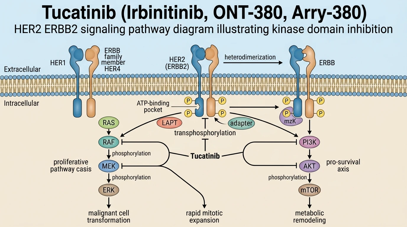
Tucatinib is a highly selective ATP-competitive inhibitor of the HER2 (ERBB2) receptor tyrosine kinase. By specifically binding the intracellular catalytic kinase domain, it blocks auto- and transphosphorylation events required for activating downstream RAS/RAF/MEK/ERK and PI3K/AKT/mTOR signaling pathways. This targeted suppression suppresses cellular mitotic activity, attenuates intratumoral hypoxia through reduced metabolic consumption, and leads to significant regression of HER2-expressing breast cancer cells while sparing EGFR-mediated pathways.
Suppression of Proliferation and Intratumoral Hypoxia Markers
Daily administration of Irbinitinib, ONT380, Arry380 induced rapid cell cycle arrest across tested models. Cellular proliferation dropped significantly within forty-eight hours of treatment initiation. Positron emission tomography utilizing [18F]-FLT showed diminished nucleoside transport and salvage pathway activity. This functional decrease closely correlated with reduced Ki-67 positivity upon histological examination. Concurrently, microenvironmental oxygenation improved markedly across treated tumors. Intratumoral hypoxia, quantified via [18F]-FMISO retention, diminished following drug exposure. Downstream nuclear accumulation of HIF-1α was attenuated in both cell line and xenograft models. Reduced cellular oxygen demand appears to alleviate metabolic strain within the core matrix.
Model-Dependent Receptor Regulation Under Kinase Blockade
Receptor tracking via [89Zr]Zr-pertuzumab PET exposed contrasting regulation patterns between experimental systems. In BT474 cell line models, surface HER2 density decreased significantly during active treatment. Intracellular kinase domain inhibition triggered down-regulation of the target receptor. However, BCM3472 patient-derived xenografts displayed no significant alteration in total target concentration. Surface binding remained constant despite evident volume regression. These divergent observations highlight model-dependent differences in receptor recycling and degradation pathways. Receptor turnover rates depend heavily on specific cell-matrix interactions and primary tissue architecture. Non-invasive immuno-PET enables precise mapping of these heterogeneous target dynamics.