GSK3326595 inhibits PRMT5 to accelerate retinal ganglion cell senescence via Wnt suppression
Copy code to add this image to your page
Glaucoma remains a primary driver of irreversible visual impairment worldwide, characterized by the progressive degeneration of retinal ganglion cells (RGCs) and corresponding axonal loss within the optic nerve head. Chronic elevation of intraocular pressure (IOP) serves as a principal mechanical stressor, inducing local tissue ischemia, microvascular compromise, and sustained cellular hypoxia. Although clinical lowering of IOP reduces disease progression in a subset of patients, neurodegenerative cascades frequently persist. Identifying intracellular signaling nodes that dictate RGC vulnerability under hypoxic and hypertensive conditions is therefore essential for developing non-IOP-dependent therapeutic strategies.
Protein arginine methyltransferase 5 (PRMT5), a type II PRMT responsible for symmetric dimethylation of arginine residues on both histone and non-histone substrates, functions as a critical regulator of chromatin architecture, transcriptional machinery, and post-translational stability. In healthy ocular tissues, PRMT5 maintains physiological gene expression profiles required for cell survival and genomic stability. However, recent quantitative expression analyses reveal that PRMT5 transcript and protein abundance are significantly downregulated in RGCs subjected to elevated IOP in animal models of glaucoma, as well as in primary RGC cultures maintained under atmospheric hypoxia (1% O2). Primary murine retinal ganglion cells isolated via immunopanning were exposed to variable durations of hypoxia. Quantitative real-time PCR and immunoblotting revealed a time-dependent loss of PRMT5 transcripts beginning as early as 12 hours post-exposure. Concurrently, nuclear localization of methyl-PRMT5 targets decreased by over 60% relative to normoxic controls. This deficit directly correlates with increased markers of premature cellular senescence, including elevated senescence-associated β-galactosidase (SA-β-gal) activity, p16INK4a and p21CIP1/WAF1 protein accumulation, and the formation of persistent γ-H2AX nuclear foci indicative of double-strand DNA breaks.
To elucidate the mechanistic consequences of decreased PRMT5 activity, pharmacologic perturbation experiments were conducted using the selective, S-adenosylmethionine (SAM)-competitive inhibitor GSK3326595 (Pemrametostat, EPZ015938). When wild-type RGCs were treated with escalating doses of GSK3326595 (0.1 to 10 µM), cellular viability dropped sharply within 48 hours. Flow cytometric analysis confirmed that this viability loss was driven predominantly by cell cycle arrest at the G1/S transition rather than acute necrotic cell death. Administration of GSK3326595 to normoxic RGC cultures phenocopied the biochemical alterations observed under chronic hypoxic stress. Specifically, treatment with GSK3326595 led to a rapid decrease in symmetric arginine dimethylation of key substrate proteins, accompanied by sustained G1 phase cell cycle arrest.
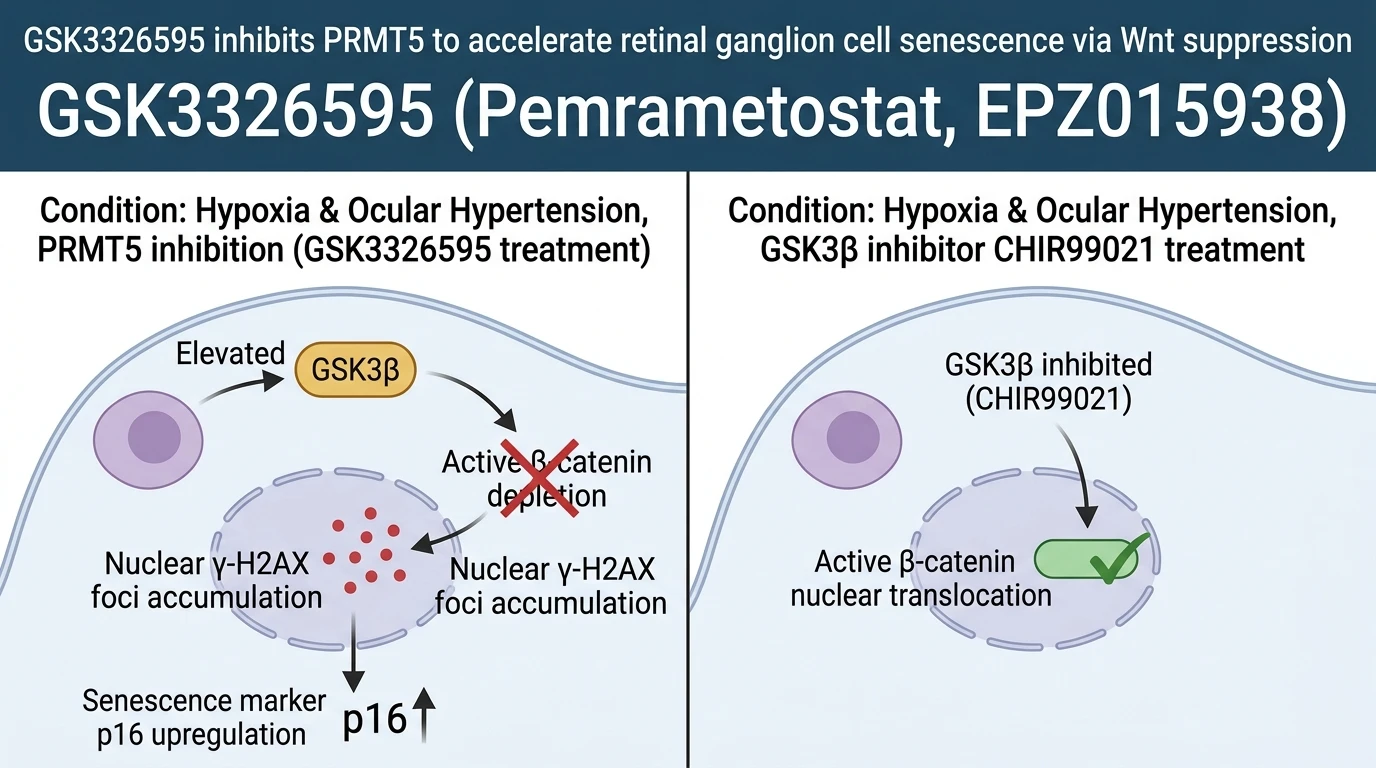
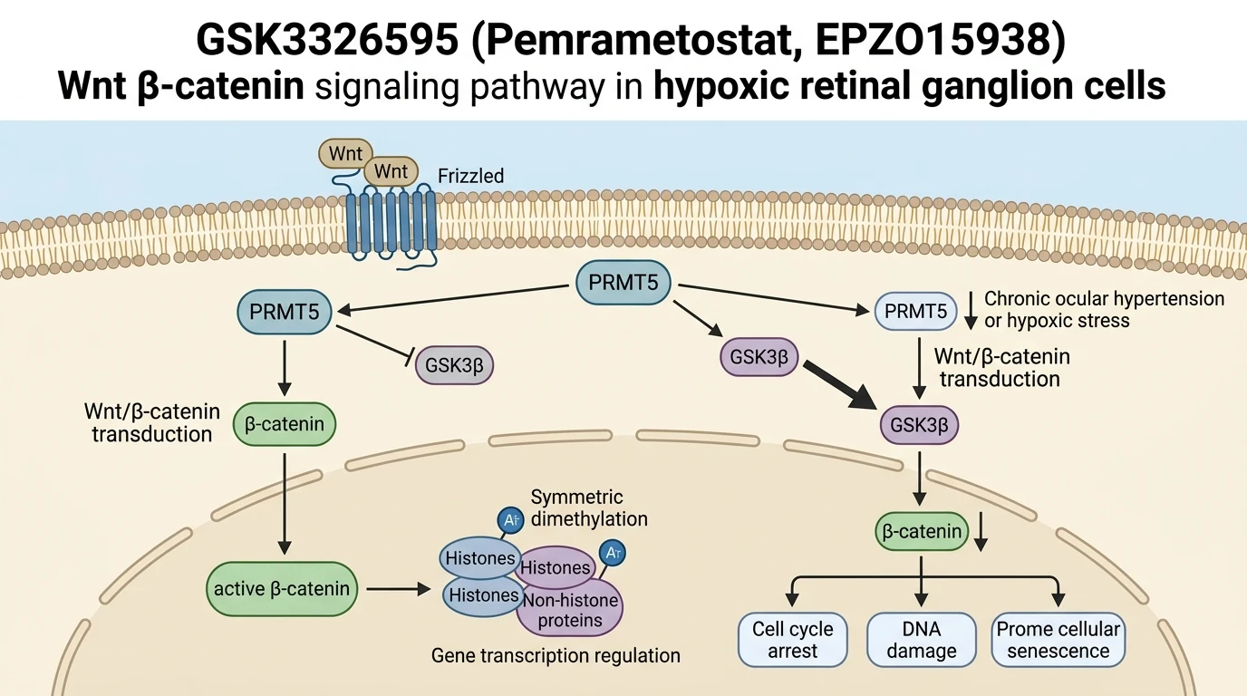
Mechanistic profiling established that PRMT5 acts as an upstream positive regulator of canonical Wnt/β-catenin signaling. Under baseline physiological conditions, PRMT5 suppresses glycogen synthase kinase 3 beta (GSK3β) expression and activity, enabling active β-catenin to accumulate and translocate to the nucleus where it drives pro-survival gene transcription. Pharmacological inhibition by GSK3326595 disrupts this axis, causing a marked elevation in total and active GSK3β levels. Consequently, active β-catenin undergoes destabilization and proteasomal degradation, terminating downstream Wnt transcriptional cascades.
In vivo intravitreal administration of GSK3326595 in rodent models markedly accelerated RGC loss and functional impairment as measured by pattern electroretinography (PERG). Retinal flat-mount immunofluorescence confirmed a profound reduction in RGC density following PRMT5 blockade. Importantly, these degenerative phenotypes were mitigated by genetic or pharmacological reactivation of the Wnt/β-catenin pathway. Adeno-associated virus (AAV)-mediated overexpression of PRMT5 in hypertensive eyes restored active β-catenin levels, suppressed GSK3β activation, reduced γ-H2AX nuclear staining, and significantly preserved RGC density. Furthermore, direct pharmacological inhibition of GSK3β using the small molecule CHIR99021 bypassed PRMT5 deficiency, downstream β-catenin signal suppression, and subsequent cellular senescence under hypoxic conditions.
These findings demonstrate that PRMT5 expression is critical for maintaining RGC survival and genomic integrity under ischemic stress. Downregulation of PRMT5 during chronic ocular hypertension destabilizes the Wnt/β-catenin signaling cascade through uninhibited GSK3β activity, accelerating RGC senescence. Target validation using GSK3326595 highlights the therapeutic potential of restoring PRMT5 activity or enforcing downstream Wnt/β-catenin activation to prevent neurodegeneration in open-angle glaucoma.
Pharmacological Evaluation and Downstream Effector Analysis
Copy code to add this image to your page
GSK3326595 acts as a potent inhibitor of Protein Arginine Methyltransferase 5 (PRMT5). In retinal ganglion cells, PRMT5 inhibition elevates GSK3β activity and suppresses active β-catenin levels, thereby turning off Wnt/β-catenin signaling. Under ocular hypertension or hypoxia, this pharmacological blockade destabilizes cell cycle homeostasis, increases genomic DNA damage, and accelerates neurodegenerative senescence in vivo.
GSK3326595 Promotes Senescence via Active β-Catenin Depletion
Targeting PRMT5 with the selective small-molecule inhibitor GSK3326595 directly established the functional requirement of arginine methyltransferase activity in maintaining retinal ganglion cell integrity. In primary RGC cultures, exposure to GSK3326595 at physiologically relevant concentrations suppressed symmetric arginine methylation in a dose-dependent manner. Active β-catenin levels dropped within 24 hours of compound exposure. This rapid loss of β-catenin destabilized nuclear transcriptional complexes, leading to cell cycle arrest at the G1/S transition. Western blot assays confirmed a concurrent elevation in total GSK3β protein—a key negative regulator of canonical Wnt signaling. In rodent eyes, intravitreal injection of GSK3326595 triggered progressive RGC somal shrinkage and optic nerve axon loss. γ-H2AX nuclear foci density increased significantly within retinal tissue mounts. These experimental observations confirm that selective PRMT5 inhibition recapitulates the cellular senescence phenotype induced by chronic ocular hypertension.
GSK3β Inhibition Restores Pathway Dynamics in Hypoxic Retinal Ganglion Cells
To confirm whether PRMT5-dependent neuroprotection operates through the canonical Wnt axis, rescue experiments were conducted in hypoxic RGC models using the GSK3β inhibitor CHIR99021. Exposure to CHIR99021 effectively blunted the accumulation of active GSK3β, maintaining cytoplasmic and nuclear pools of active β-catenin despite PRMT5 downregulation. In hypoxic cell assays, CHIR99021 co-treatment reduced SA-β-gal positive cell fractions by 45% compared to vehicle controls. Expression of downstream target genes, including cyclin D1 and c-Myc, was maintained at physiological levels. Similarly, AAV-mediated restoration of PRMT5 expression in hypertensive murine eyes sustained RGC survival rates over a four-week monitoring period. Electroretinographic amplitudes remained preserved relative to sham-treated controls. These empirical findings demonstrate that PRMT5 functions as an essential upstream regulator of Wnt/β-catenin signaling. Re-establishing this signaling pathway offers a viable pharmacological strategy to attenuate retinal ganglion cell senescence under ischemic stress conditions.